GENERAL MEDICINE
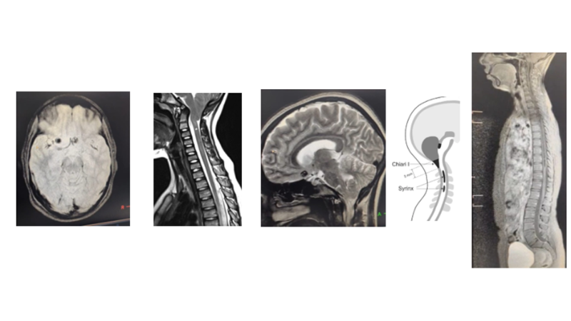
- A 34-year-old woman presented with 2 months of brief staring spells, chronic headache and intermittent fever. MRI showed neurocysticercosis with incidental Chiari I malformation and syringomyelia. She was treated with albendazole, steroids and sodium valproate, leading to clinical improvement and stable discharge with follow-up. No focal deficits were noted clinically.
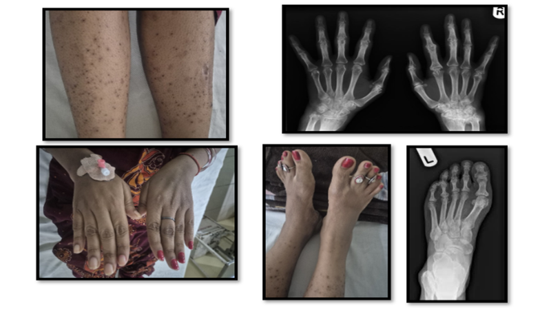
- A 33-year-old woman presented with polyarthritis, fever, rash, and edema. Evaluation confirmed rheumatoid arthritis with Sjögren’s syndrome and vasculitic complications. Labs showed anemia, leukocytosis, elevated ESR, and positive ANA and autoantibodies. Treated with NSAIDs and tofacitinib, she improved significantly, regained function, and resumed normal daily activities.
GENERAL SURGERY
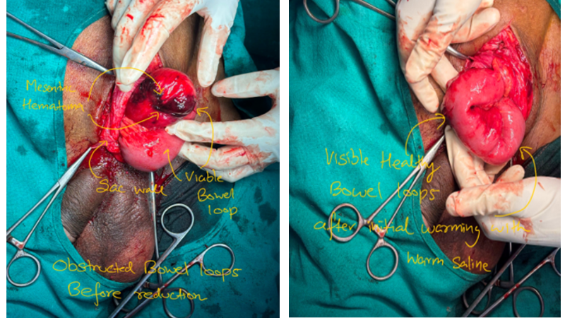
- A 60-year-old man presented with obstructed left inguinal hernia. Emergency surgery revealed viable congested bowel with mesenteric hematoma. Adhesiolysis, reduction, and herniorrhaphy were performed without resection; hydrocele sac eversion was added. Postoperatively stable, he passed flatus and stools, tolerated oral intake, highlighting timely management and effective surgical care.
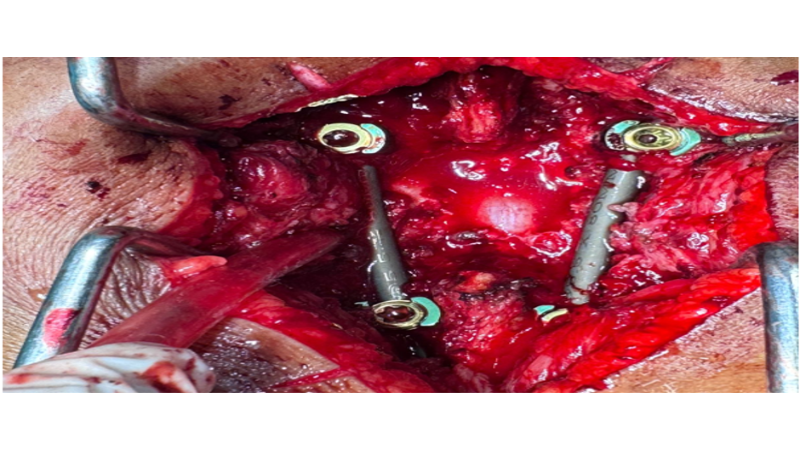
- A 39-year-old woman presented with progressive low back and bilateral leg pain for three months. Initially treated conservatively and empirically for spinal tuberculosis without improvement, MRI was advised. Neurological exam showed L4–L5 sensory deficits without motor weakness. Case referred for specialist evaluation and differential diagnosis due to worsening symptoms despite therapy.
RADIOLOGY DEPARTMENT
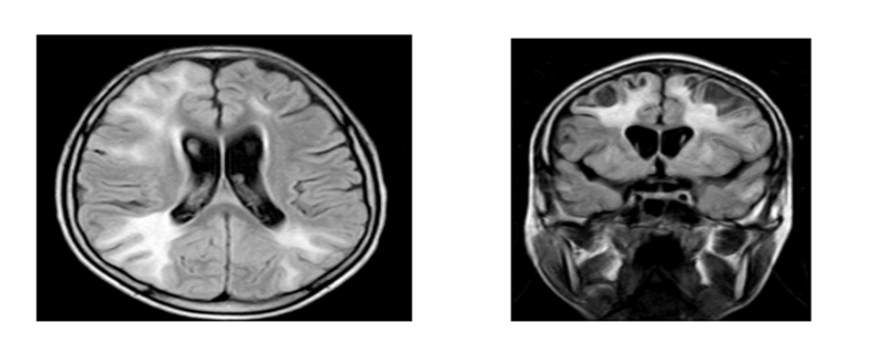
- MRI in Acute Disseminated Encephalomyelitis (ADEM)
MRI is the imaging modality of choice for diagnosis and follow-up. Usually occurs 1–3 weeks after viral infection or vaccination, commonly in children. Multifocal, asymmetric, poorly defined demyelinating lesions. Lesions are T2/FLAIR hyperintense and T1 hypointense. Predominantly involve subcortical and deep white matter. Thalamic and basal ganglia involvement is common, favouring ADEM over multiple sclerosis. May involve the brainstem, cerebellum, corpus callosum, and spinal cord. Variable contrast enhancement (patchy, ring, or open-ring); mild edema/mass effect may occur. Spinal cord: Long-segment T2 hyperintensity. Follow-up MRI: Partial/complete lesion resolution; persistent or new lesions suggest multiple sclerosis or MOG antibody-associated disease (MOGAD).
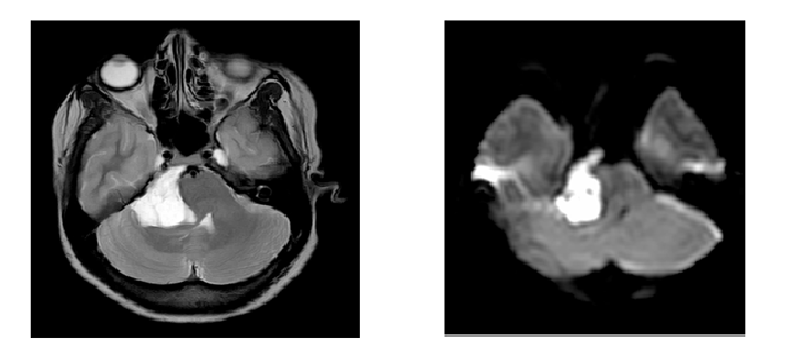
- MRI in Epidermoid Cyst - Second most common CPA mass after vestibular schwannoma. Congenital ectodermal inclusion cyst; MRI is the investigation of choice. Common sites: Cerebellopontine angle (40–50%), parasellar cistern, and fourth ventricle. T1: Hypointense; T2: Hyperintense (CSF-like). FLAIR: Incomplete CSF suppression (dirty CSF sign). DWI: Marked diffusion restriction (most characteristic feature); low ADC. Post-contrast: No enhancement (rare thin peripheral enhancement). Morphology: Lobulated extra-axial lesion with cauliflower margins, encasing vessels and cranial nerves, with cisternal insinuation. Differentials: Arachnoid cyst (no diffusion restriction, complete FLAIR suppression) and dermoid cyst (T1 hyperintense)
ENT DEPARTMENT
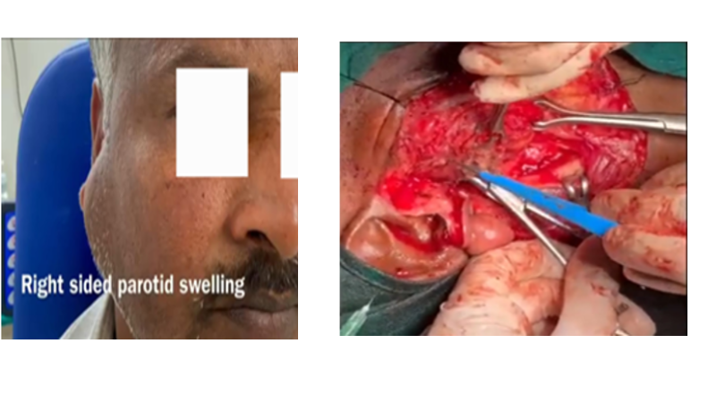
- Superficial parotidectomy is performed for benign tumours involving the superficial lobe of the parotid gland, commonly pleomorphic adenoma. The procedure requires meticulous identification, dissection and preservation of the facial nerve and all its branches while achieving complete tumour excision. It is technically demanding because of the complex anatomy and the risk of facial nerve injury. Careful surgical technique minimises complications such as facial weakness, Frey syndrome and salivary fistula while ensuring good oncological clearance and satisfactory cosmetic and functional outcomes.
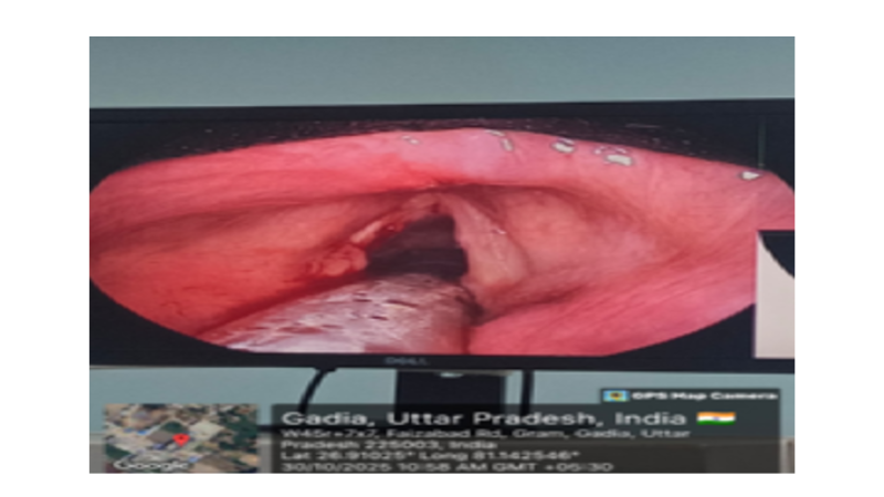
- Microlaryngoscopy is a specialised endoscopic procedure performed under general anaesthesia for diagnosis and management of laryngeal disorders. Using an operating microscope and fine microsurgical instruments, lesions such as vocal cord polyps, nodules, cysts and suspicious growths are precisely excised or biopsied while preserving normal vocal fold tissue. The procedure demands excellent hand-eye coordination, magnified visualisation and meticulous tissue handling to maintain postoperative voice quality. It is one of the most technically demanding endolaryngeal surgeries and plays an important role in the diagnosis and treatment of benign and early malignant laryngeal lesions.
OPTHALMOLOGIST DEPARTMENT
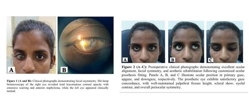
- A 23-year-old woman presented with a disfigured, blind right eye secondary to anterior staphyloma, total corneal opacity, and advanced corneal degeneration. Visual acuity was absent light perception in the right eye, while the left eye had normal vision. She sought cosmetic rehabilitation with a customized ocular prosthesis to restore facial symmetry and confidence. Following comprehensive ocular and systemic evaluation, uncomplicated right-eye evisceration was performed. The postoperative course was satisfactory, with healing and formation of a stable, well shaped socket. Three months later, she was fitted with a customized ocular prosthesis, achieving excellent anatomical alignment, facial symmetry, and a pleasing aesthetic outcome.
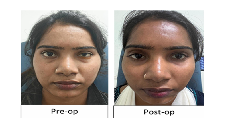
- A 24-year-old woman presented with mild congenital ptosis of the left upper eyelid, causing noticeable facial asymmetry and reduced confidence in her workplace. A comprehensive ophthalmic evaluation was performed, including assessment of visual acuity, eyelid position, levator function, ocular motility, and protective mechanisms. Based on the clinical findings, ptosis correction using the Fasanella Servat procedure was planned and performed successfully. The postoperative recovery was uneventful, with satisfactory elevation and contour of the left upper eyelid. At follow-up, excellent eyelid symmetry, appropriate palpebral fissure height, and preserved ocular function were achieved, resulting in a pleasing aesthetic outcome and improved her workplace confidence.